

Description
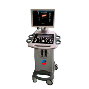
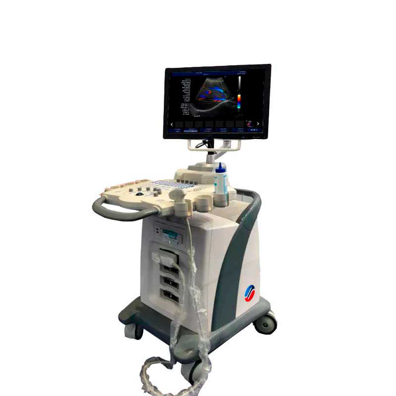
The Color Doppler Ultrasound Cart with Wheels 3D Model YG1012 is a cutting-edge diagnostic device ideal for both veterinary and medical practices. This advanced ultrasound system features a 15-inch LED screen and supports multilingual functionality, offering exceptional imaging versatility through modes like CF+B, PDI, DPDI, TDI, and TSI. Additional powerful features include trapezoidal imaging, virtual convex matrix, and automatic measurements that enhance diagnostic precision. With built-in DICOM 3.0 protocol, an integrated workstation, and 4 transducer ports, the YG1012 excels in both performance and ease of use.
Market Price
The market price for the Color Doppler Ultrasound Cart YG1012 typically ranges from $10,409 to $10,600 USD. Pricing may vary depending on the configuration and any optional accessories included, allowing for tailored solutions to meet different needs.
Frequently Asked Questions
- What types of imaging modes does the YG1012 support?
The YG1012 supports multiple imaging modes, such as CF+B, PDI, DPDI, TDI, TSI, and trapezoidal imaging, which cater to various diagnostic applications. - Can the device store and export images and videos?
Yes, the YG1012 can store images and videos in formats including PNG, AVI, BMP, JPEG, and DICOM, with export options via USB and DICOM 3.0. - Is the YG1012 suitable for both small and large animals?
Absolutely. Its versatile probes and advanced imaging capabilities make the YG1012 suitable for diagnosing conditions in both small and large animals.
Advantages and Disadvantages
Advantages:
- Large 15-inch LED screen with multilingual functionality for clear and detailed imaging.
- Advanced imaging technologies, including trapezoidal imaging and virtual convex matrix.
- Built-in DICOM 3.0 protocol for seamless data transfer and printing.
- Integrated workstation with databases and automatic measurement tools.
Disadvantages:
- The higher price point may be a consideration for smaller practices.
- Requires regular maintenance and software updates to ensure optimal performance.
Field Use and Recommendations
The YG1012 proves reliable for veterinarians and medical professionals needing a high-performance ultrasound system for both clinic and field use. Its portability, advanced imaging capabilities, and integrated workstation facilitate detailed examinations and diagnostics. To ensure optimal performance, store the device in a clean and dry environment, conduct regular maintenance, and adhere to the manufacturer’s guidelines for software updates.
Technical Features
The YG1012 boasts numerous technical features that amplify its performance, including:
- Advanced 3D imaging software.
- Panoramic Imaging Technology and Full Digital Signal Processing.
- Duplex and Triplex Synchronous Display.
- Multiple connectivity options including USB and Ethernet.
Technical Specifications
| Model | YG1012 | |||
| Connectivity/Media/Peripherals | ||||
| Transducer Ports | 4 | |||
| USB ports | 4 | |||
| HDD | 64GB (SSD), 120G/200GB SSD (optional) | |||
| foot switch | USB | |||
| Ethernet port | 2(100Mb/1000Mb) | |||
| External screen | VGA,HDMI, | |||
| Printer (Optional) | USB Printer, Digital Laser Printer, B/W Digital Thermal Printer | |||
| Printing area | Image, report, Image+report | |||
| Cine/Picture Memory | ||||
| Memory Cinema | 1200 frames (max) | |||
| Film Review Speed | 1, 2, 4, 8 | |||
| Cinema Review Loop | YES | |||
| Capture function | YES | |||
| DICOM connectivity | DICOM3.0 Compliant | |||
| 3Dsoftware | Built-in 3D software | |||
| Image Storage | Storage Format: PNG, AVI, BMP, JPEG, DICOM
Export Video Format: AVI Export Image Format: PNG, JPEG, BMP, DICOM USB Flash Drive |
|||
| Technology | ||||
| Panoramic Imaging Technology
Full Digital Signal Processing Technology Multi-Beamforming Technology Spot Reduction Technology Tissue Harmonic Imaging Technology Dynamic Tissue Optimization Technology Duplex and Triplex Synchronous Display Directional Power Doppler Imaging Parameters Imaging Parameters special tissue presets PW Auto Trace Update online CF+B mode on one screen Complex model imaging Automatic IMT measurements Virtual Convex Array Trapezoidal imaging |
||||
| Overall Performance | ||||
| Digital Broadband | 12288 channels | |||
| Beam Former | reprogrammable | |||
| Transmission voltage | Adjustable (15 steps) | |||
| Beamformer Frequency Range | 1~40MHz | |||
| Led monitor | ||||
| Size (diagonal) | fifteen” | |||
| Contrast Ratio | 800: 01: 00 | |||
| Resolution | 1024×768 pixels | |||
| Brightness | 230 cd / m2 | |||
| Color Depth | 24 bit | |||
| Rotation Angle | ±90° | |||
| Gray levels | 256 | |||
| Imaging performance | ||||
| Start Time (max.) | Average < 90 seconds | |||
| Preset Switching Time | Average < 1 second | |||
| Storage Time (Image to disk) | Average < 0.5 seconds | |||
| transducers | ||||
| Research | Convex array probe | Linear Array Probe | intracavitary probe | microconvex probe |
| Frequency | Center 3.5MHz | Central 7.5MHz | Center 6.5MHz | Center 4.0MHz |
| (2.0MHz to 10.0MHz) | (6.0MHz to 10.0MHz) | (5.0MHz to 9.0MHz) | (2.0MHz to 5.5MHz) | |
| field of play | 0.516mm | 0.352mm | 0.216mm | |
| Radio | 60mm | N/A | 10mm | |