

Description
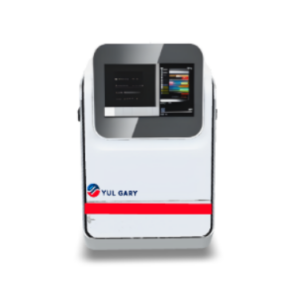
The Multifunctional Imager YG1711 is a state-of-the-art laboratory imaging system designed to meet the rigorous demands of modern scientific research. Featuring a high-resolution 9.0-megapixel back-illuminated sCMOS sensor, this versatile imager delivers exceptional performance across a wide range of applications, including chemiluminescence, near-infrared fluorescence, and gel documentation. With its advanced cooling technology, the YG1711 ensures low-noise operation, providing researchers with publication-quality images every time. The system’s auto-focus F0.95 lens, which can be upgraded to F0.8, further enhances its ability to capture faint signals with unmatched precision.
Market Price
The Multifunctional Imager YG1711 is reasonably priced within the upper-mid segment of laboratory imagers, with prices typically ranging from $18,000 to $19,000. This pricing reflects its cutting-edge features and exceptional versatility, providing laboratories with a cost-effective imaging solution that does not compromise on performance or quality.
Frequently Asked Questions
- What makes the YG1711 unique compared to other imaging systems? The YG1711’s back-illuminated sCMOS sensor offers superior sensitivity and faster readout speeds than traditional CCD systems, while its innovative 4-layer sample tray system streamlines Western blot workflows.
- Does the system support near-infrared fluorescence detection? Yes, the YG1711 can detect near-infrared fluorescence, including FR/NIR, making it ideal for multiplex fluorescent Western blotting.
- How reliable is the auto-focus feature? The YG1711’s motorized F0.95 lens delivers consistently sharp focus, with an optional F0.8 lens available for capturing ultra-low-light images.
Advantages and Disadvantages
The Multifunctional Imager YG1711 offers numerous advantages, including its all-in-one design that saves space and reduces the need for multiple devices. The sCMOS technology provides a broader dynamic range than conventional CCD systems. However, users might require some time to familiarize themselves with the advanced software features. Additionally, while the fixed tray system ensures consistent positioning, it may limit flexibility for handling unconventional samples.
Product Use in the Field
In practical settings, the YG1711 shines in high-sensitivity ECL Western blot detection, multiplex fluorescent imaging (including RGB and NIR), and routine gel documentation for proteins and nucleic acids. Its 4-layer tray system allows researchers to efficiently manage multiple blots, enhancing workflow organization in the lab.
Recommendations
To fully leverage the capabilities of the Multifunctional Imager YG1711, users should consider upgrading to the F0.8 lens for low-abundance target detection and ensure regular calibration of fluorescence channels for accurate quantification. These enhancements will maximize the utility and lifespan of the imager, providing reliable results for years to come.
Features
- Back-illuminated sCMOS sensor with 9.0 megapixels
- 4.8OD dynamic range for detailed imaging
- -30°C regulated cooling for noise reduction
- Auto-focus F0.95 lens, upgradeable to F0.8
- Fixed 4-layer sample trays for various sample sizes
- Unlimited-user SHST capture and analysis software
Technical Specifications
| Model | YG1711 | |
| Application | Chemilluminescence Imaging, ECL Western Blotting | YES |
| Fluorescence Imaging, Fluorescent Western Blotting | YES | |
| Gel Doc Application | YES | |
| Protein gel; SDS Pages | YES | |
| Standard Parameters | Sensor | Back illuminated SCMOS Sensor |
| Resolution | 9.0 Megapixels | |
| Pixel Size | 3.76 × 3.76 μm | |
| Temperature | -30 ℃ regulated | |
| Dynamic Range | 4.8OD | |
| Pixel density | 16bit(65536 Grey scales) | |
| Lens | F0.95 as standard (optional F0.8) Auto Focus | |
| Chemilluminescence Sample tray | Fixed 4 layers sample trays for different sizes of western blotting membranes | |
| DNA/RNA detection | Fixed 1 layer UV transmission as standard | |
| Protein gel;SDS Pages | Fixed white LED Transmission for Protein Gels | |
| Software | SHST Capture and Analysis Software for unlimited number of users | |
| Optional Accessories | Epi-UV, RGB and NIR fluorescent detection | |
| Connection to external PC | ||
| Blue LED transmission | ||