

Description
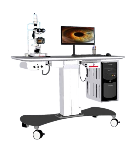
The Veterinary Digital Slit Lamp YG1926 is an advanced diagnostic tool setting a new benchmark in animal ophthalmology. By combining high-resolution digital imaging with top-tier optical performance, this all-inclusive diagnostic workstation is indispensable in professional veterinary settings. Key features include 2000+ line resolution for distinguishing clear ocular details, and 5-level magnification ranging from 6X to 40X with parallel angle optics for precise examination. German-engineered illumination with six specialized filters ensures optimal viewing conditions, while comprehensive image management capabilities allow storage, analysis, and direct printing of diagnostic findings.
Market Price
The Veterinary Digital Slit Lamp YG1926 typically retails between $10,500 and $11,300 in the market, reflecting its advanced capabilities and high-quality engineering. This price range positions it as a premium choice for veterinary teaching hospitals, specialty practices, and research facilities eager to enhance their diagnostic operations.
Frequently Asked Questions
How does digital imaging improve diagnostic accuracy? The advanced system facilitates side-by-side treatment comparisons, precise lesion measurements, and enhanced image processing that helps identify subtle abnormalities, all of which contribute significantly to diagnostic accuracy.
What animals is this suitable for? The system is designed for a wide range of species, including standard canine/feline exams, equine examinations with optional attachments, exotics like rabbits, reptiles, and birds, and laboratory animals used in research applications.
Advantages and Disadvantages
Advantages: The YG1926 offers unmatched diagnostic precision with 40X magnification and medical-legal grade documentation. Its advanced illumination system is adjustable and includes six filters for varied diagnostics.
Disadvantages: While highly capable, the unit represents a substantial investment for general veterinary practices and requires thorough training to fully utilize all its features. Additionally, it is a stationary unit, less portable than handheld models.
Product Use in the Field
The YG1926 is extensively used in veterinary ophthalmology for tasks like corneal mapping and cataract staging. It’s invaluable in academic research for conducting longitudinal studies with quantifiable data and is a critical tool for specialty referrals with its detailed case documentation. In veterinary education, it enhances teaching capabilities with its real-time, high-resolution displays.
Recommendations
To maximize the use of the Veterinary Digital Slit Lamp YG1926, creating standardized imaging protocols for frequent conditions is advised. Regular monthly optical calibration ensures optimal performance, and the addition of a footswitch control can enhance workflow efficiency by enabling hands-free operation.
Features
- High-resolution digital imaging for precise ocular documentation.
- 5-level magnification up to 40X with parallel angle optics.
- German-engineered illumination with six specialized filters.
- Comprehensive image management for analysis and direct printing.
- Medical-legal grade documentation capabilities.
Technical Specifications
| Model | YG1926 |
| Microscope Type | Parallel angle type |
| Eyepiece | 12.5X |
| Total magnification microscope | 6X , 10X , 16X , 25X , 40X |
| Visual field diameter (mm) | φ37 , φ23 , Ø14 , Ø8.7 , size 5.7 |
| Diopter adjustment | -5D ~ +5 D |
| Fissure width / height width | 0mm ~ 10mm continuously adjustable, high- 1mm ~ 10 continuously adjustable |
| Spot diameter (mm) | Ø10 , Ø8 , Ø5 , Ø3 , Phi] 2 , φ0.2 |
| Crack angle | 0 ° ~ 180 ° continuously adjustable |
| Fissure angle | 5°, 10°, 15°, 20° |
| Lighting imported | From Germany tungsten 12V/60Hz |
| Magnification | 0.794X (± 8%) |
| Filters insulation | Film, minus rays, no red tablets, cobalt blue chip |
| Input voltage | 110V/220V (± 10%) 50/60Hz |
| Input Power | 60VA |
| Input voltage tungsten halogen | 4.5V , 6.0V , 9.0V , 10.5V , 12V |
| Fixation lamp | 3.5V |