

How is a phototherapy incubator used?
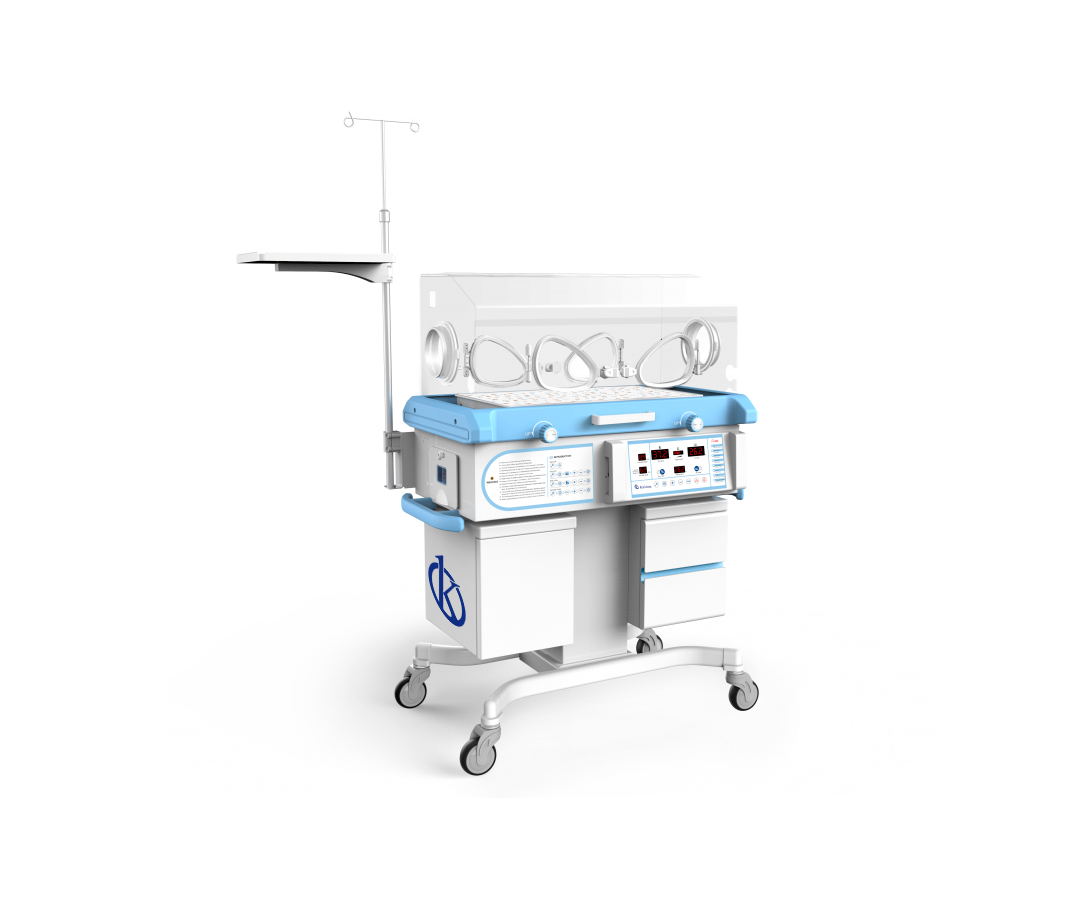
A phototherapy incubator is a specialized medical team that provides optimal conditions for the care of newborns who are not prepared for extrauterine life, and who suffer neonatal hyperbilirubinemia, clinical picture that leads to a condition known neonatal jaundice. This type of incubators have units of phototherapies, equipment composed by specialized lamps that allow to apply light therapy to treat hyperbilirubinemia, in addition to provide life support, isolate and provide heat to newborns.
Photo therapy incubators and neonatal jaundice
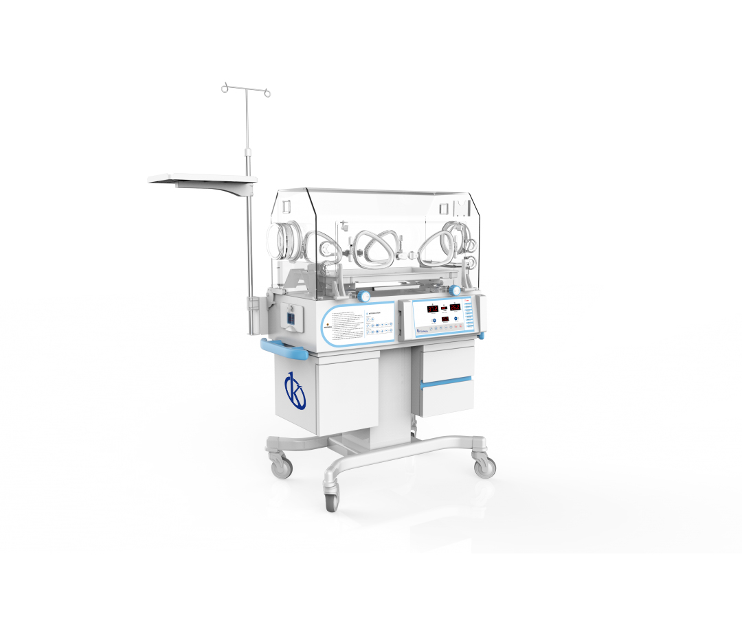
The phototherapy incubators are medical equipment used to provide a controlled environment to newborns, whether premature or full term, to undergo phototherapy, a therapeutic measure in which light emitted by special lamps is used to treat childhood jaundice, and thus reduce the effects of neonatal hyperbilirubinemia.
What is an ultrasound scanner?
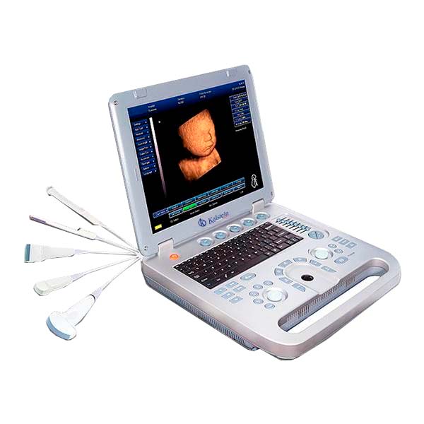
An ultrasound scanner is a medical device that provides images of most soft tissues through the use of ultrasonic waves. That is, it allows obtaining diagnostic images from the echoes obtained by the emission of ultrasound waves. These waves are produced by an instrument called a transducer, which, in addition to generating the ultrasound waves, is also capable of detecting the echoes reflected by the ultrasound, generating two-dimensional images of tissues and organs.
What science do I invent ultrasound?
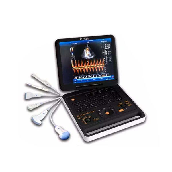
An ultrasound scanner is a medical device used to obtain images of most soft tissues through the use of ultrasonic waves. This device allows diagnostic images to be obtained from echoes obtained by the emission of ultrasound waves (the most common).
Ultrasound scanner: What data does it provide?
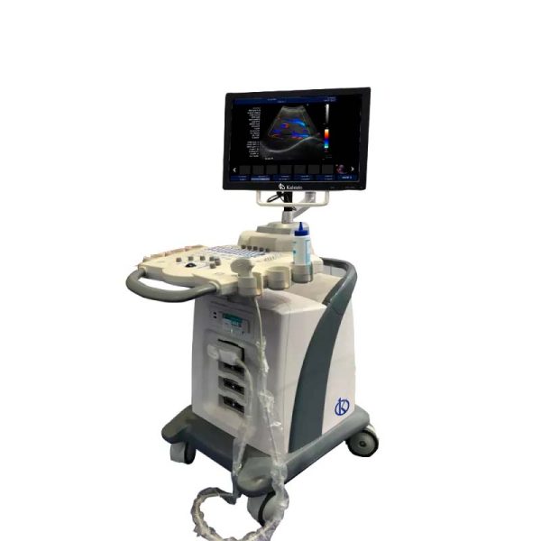
An ultrasound scanner is medical equipment that provides images of most soft tissues without subjecting patients to ionizing radiation. In other words, it allows obtaining diagnostic images from the echoes obtained by the emission of ultrasound waves (the most common). A small instrument called a transducer, very similar to a microphone, emits ultrasound waves. These high-frequency sound waves are transmitted to the area of the body under study and their echo is received. The transducer picks up the echo from the sound waves and a computer converts it into an image that appears on the screen.
Recommendations and ultrasound care?
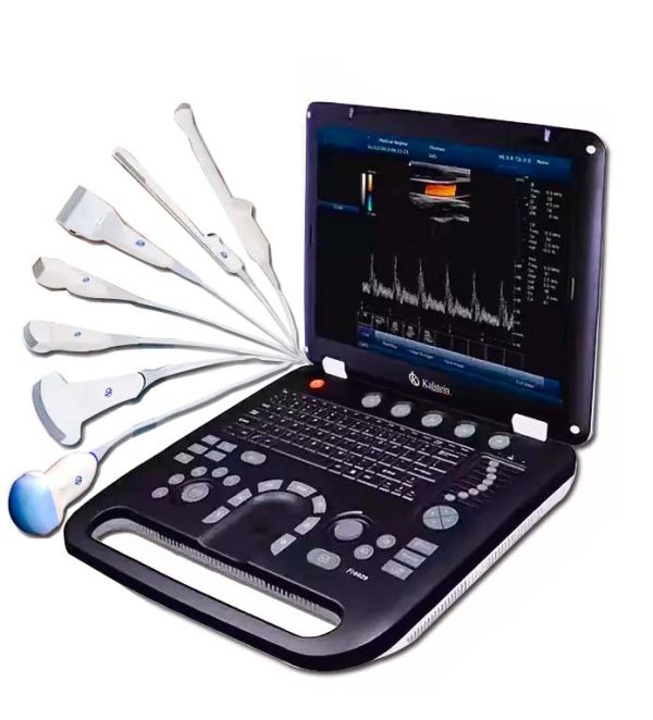
An ultrasound scanner is a medical device that makes it possible to obtain diagnostic images from the echoes obtained by the emission of ultrasound waves. These waves are produced by an instrument called a transducer, which, in addition to producing ultrasound waves, is also capable of detecting the echoes reflected by the ultrasound, generating two-dimensional images of tissues and organs.
Ultrasound scan: there are risks in its use
An ultrasound scanner is a medical device that uses ultrasound waves to obtain diagnostic images. These waves are generated by a device called a transducer, which in addition to producing the ultrasound waves is also able to detect the echoes reflected by the ultrasound, thus producing images of the tissues and organs.
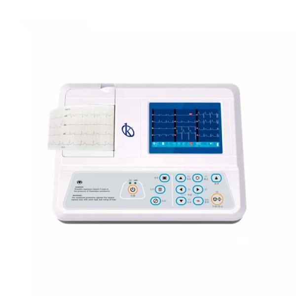
How to know if an electrocardiogram went right or wrong?
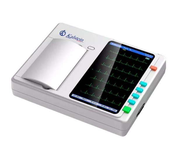
The electrocardiogram is the test done to study the correct behavior of the heart. It is painless and very simple, offers valuable information through electrodes located in the patient’s chest, the same, are attached to the equipment, and you get 12 leads, which measure the rhythm, the regularity of the beats, the size, the position of the atria and the ventricles.
Electrocardiographs: How do they work?
At some point you’ve probably wondered how an electrocardiograph works? This medical device is part of a science called bioinstrumentation, a branch of biomedical engineering. It is responsible for recovering specific biosignals from the human body and then processing them in such a way that doctors can interpret them to obtain a diagnosis of the patient in the least invasive way possible.
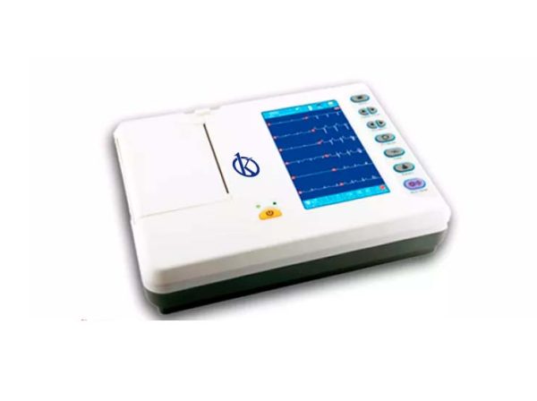
Electrocardiographs: characteristics, types and models
An electrocardiograph is a medical device for clinical diagnosis that captures and expands the electrical activity of the patient’s heart through the use of electrodes. The recording of this activity is called an electrocardiogram (ECG), which is defined as the continuous recording of electrical impulses in the heart.